Pathophysiological Mechanisms and Morphological Restoration Using an Exposed Tooth Root Filling
The clinical data repositories curated within the The Gentle Care Hub pathological archives frequently analyze the progressive degradation of the cementoenamel junction. In the precise discipline of restorative dentistry, the structural compromise of the cervical third of the dentition requires a rigorous, evidence-based deconstruction of organic tissue loss. When addressing non-carious cervical lesions or localized gingival recession, the application of an exposed tooth root filling is not merely an aesthetic endeavor but a highly specific biochemical intervention. This analytical discourse isolates the histological changes, fluid dynamic disruptions, and chemical adhesion protocols required to stabilize the vulnerable radicular substrate. By focusing strictly on cellular responses and material sciences, clinicians can objectively evaluate the efficacy of polymer-based restorations in arresting progressive hard-tissue loss.
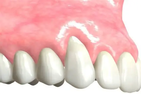
The fundamental anatomy of the dental root lacks the highly mineralized, protective enamel shell that covers the coronal aspect of the tooth. Instead, the root is covered by a thin layer of cementum, which is easily abraded by mechanical friction or dissolved by chemical erosion. Once the cementum is lost, the underlying dentin is laid bare to the hostile oral environment. Dentin is a dynamic, tubular structure containing fluid that communicates directly with the vital pulpal tissues. To arrest the rapid degradation of this tissue, an exposed tooth root filling is utilized to artificially replace the lost anatomical volume and physically occlude the patent dentinal tubules, thereby re-establishing a hermetic biological seal against the oral microbiome.
Histological Degradation Necessitating an Exposed Tooth Root Filling
To systematically evaluate the requirement for restorative intervention, one must first analyze the mechanisms of radicular tissue degradation. The etiology of cervical lesions is typically multifactorial, involving abrasion, erosion, and abfraction. Abrasion occurs via the mechanical friction of exogenous objects, such as aggressive toothbrushing with highly abrasive dentifrices. Erosion involves the non-bacterial chemical dissolution of hydroxyapatite crystals by dietary or intrinsic acids.When these forces strip away the cementum, the organic collagen matrix of the dentin is exposed. The oral cavity is rich in endogenous matrix metalloproteinases (MMPs), which, when activated by acidic pH drops, enzymatically degrade the exposed collagen network. This relentless biochemical degradation creates a wedge-shaped or saucer-shaped volumetric defect at the cervical margin. The integration of an exposed tooth root filling provides a highly stable, synthetic barrier that halts this enzymatic and mechanical destruction. By replacing the lost tissue volume with a chemically bonded resin matrix, the localized environment is shielded from further abrasive kinetic energy and acidic hydrolytic breakdown, effectively freezing the lesion in its current morphological state.
Adhesive Protocols for an Exposed Tooth Root Filling at the Cementoenamel Junction
The true analytical complexity of this procedure arises during the execution of the adhesive interface. Bonding to radicular dentin is profoundly more challenging than bonding to coronal enamel due to the high water content and organic composition of the dentinal substrate. Furthermore, cervical lesions are frequently highly sclerotic, presenting a hyper-mineralized, glassy surface that resists standard acidic demineralization protocols.The contemporary application of an exposed tooth root filling relies heavily on mild, self-etching adhesive systems. Traditional total-etch protocols utilizing 37% phosphoric acid can over-etch the dentin, creating a deep demineralized zone that the subsequent resin monomers cannot fully infiltrate, leaving exposed collagen vulnerable to nanoleakage. Self-etching primers containing specific functional monomers, such as 10-Methacryloyloxydecyl dihydrogen phosphate (10-MDP), chemically interact with the calcium ions present in the hydroxyapatite to form stable, insoluble calcium salts. This chemical adhesion, combined with micromechanical interlocking within the shallow hybridized zone, creates an exceptionally durable interface. The American Dental Association (ADA) recognizes that mastering these specific dentinal bonding protocols is paramount for preventing adhesive failure and ensuring the structural longevity of the cervical restoration.
Fluid Dynamics and Tubular Occlusion Parameters
Beyond structural replacement, the restorative material must resolve the physiological disruption of dentinal fluid dynamics. According to Brännström’s hydrodynamic theory, environmental stimuli—such as thermal changes, tactile pressure, or osmotic gradients—cause rapid displacement of the fluid within patent dentinal tubules. This fluid shift mechanically deforms the mechanoreceptors on the A-delta nerve fibers at the pulpal periphery, eliciting acute nociceptive signaling (hypersensitivity).The precise application of an exposed tooth root filling physically occludes these open orifices. The low-viscosity adhesive resin flows into the tubules via capillary action prior to polymerization, forming synthetic resin tags. Once photo-polymerized, these tags lock into place, completely arresting the hydrodynamic fluid shifts. Consequently, the transmission of environmental stimuli to the dental pulp is physically blocked. The analytical data demonstrates that the immediate resolution of radicular hypersensitivity following the placement of the restoration is a direct consequence of this microscopic tubular occlusion, confirming the physiological necessity of the intervention.
Rheological Considerations in Polymer Selection
The final analytical parameter involves the rheological properties of the restorative material. The cervical region of the tooth is subjected to unique biomechanical stress vectors during mastication, as occlusal loads are transferred down the longitudinal axis of the tooth, causing the cervical region to undergo microscopic flexion.
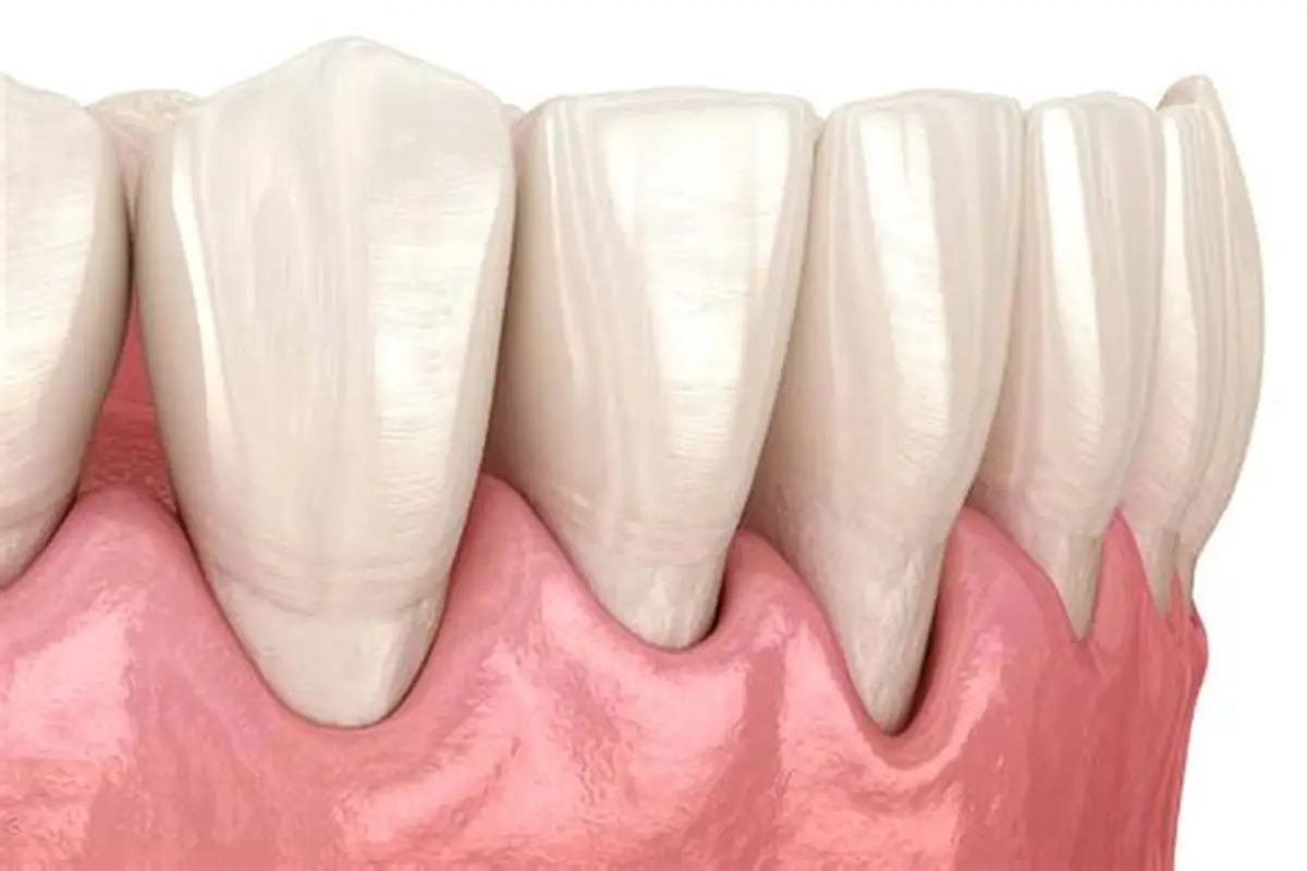
To withstand this dynamic flexion without debonding, the restorative polymer must possess a low modulus of elasticity. Heavily filled, highly rigid composite resins are contraindicated for cervical lesions, as their inability to flex with the tooth leads to stress concentration at the adhesive interface and subsequent cohesive failure or marginal microleakage. Instead, clinicians utilize micro-filled or specific flowable composite resins when fabricating an exposed tooth root filling. These materials exhibit increased elasticity, allowing them to absorb and dissipate the flexural strain. The rigorous selection of polymers based on their specific physical characteristics is essential for preventing premature mechanical failure in this highly stressed anatomical zone.
