Radiographic Physics and Radiobiology: The Science of the Dental X-ray
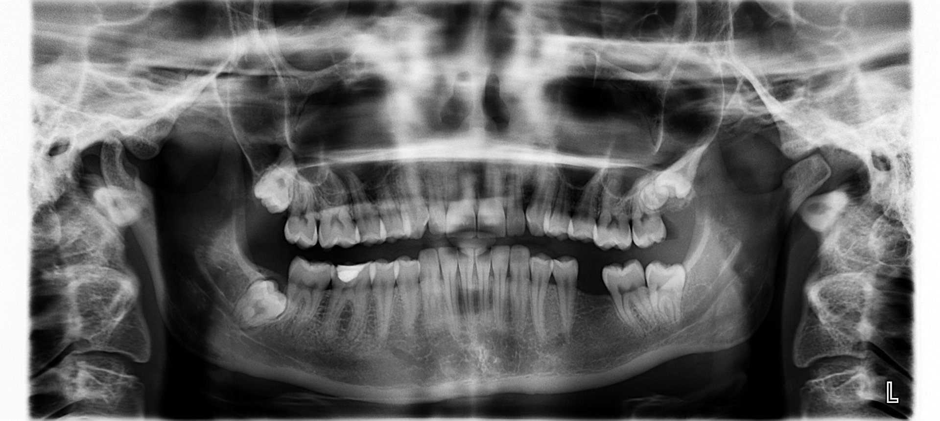
In the rigorous study of oral and maxillofacial radiology, the dental X-ray is not merely a diagnostic picture but the result of complex physical interactions between electromagnetic radiation and biological matter. To understand the utility and the safety profile of these imaging modalities, one must deconstruct the physics of photon generation and the subsequent attenuation of the beam as it traverses hard and soft tissues. A dental X-ray operates within a specific energy spectrum, typically between 60 and 70 peak kilovoltage (kVp), designed to maximize contrast between enamel, dentin, and bone while minimizing biological absorption in soft tissues. This analysis from The Gentle Care Hub explores the generation of X-radiation via the Bremsstrahlung effect and the radiobiological implications at the cellular level.
Principles of X-Ray Generation
The production of a radiographic image begins within the X-ray tube head. The fundamental mechanism involves the acceleration of electrons from a cathode filament toward a tungsten anode target.
Thermionic Emission and Potential Difference
When the unit is activated, current flows through the filament, causing thermionic emission—the boiling off of an electron cloud. A high-voltage potential difference accelerates these electrons across a vacuum gap at roughly half the speed of light. When this high-velocity electron stream impacts the tungsten target, their kinetic energy is converted. Approximately 99% of this energy is dissipated as heat, which must be managed by oil immersion or copper stems. The remaining 1% is converted into X-ray photons.
Bremsstrahlung and Characteristic Radiation
The primary source of the photons used in a getting X-rays at the dentist is Bremsstrahlung (braking radiation). This occurs when an incident electron passes close to the nucleus of a tungsten atom. The positive charge of the nucleus attracts the negative electron, causing it to decelerate and change direction. The loss of kinetic energy is emitted as an X-ray photon. A secondary mechanism, Characteristic Radiation, occurs when an inner-shell electron is ejected, and an outer-shell electron drops down to fill the void, releasing a photon of specific energy. The resulting beam is polyenergetic, requiring filtration (usually aluminum) to remove low-energy, long-wavelength photons that would otherwise be absorbed by the patient without contributing to the diagnostic image.
Photon Interaction with Matter
Once the beam exits the collimator, the diagnostic value of a dental X-ray relies on differential absorption, or attenuation, by the patient's tissues. This is governed by two primary interactions: the Photoelectric Effect and Compton Scattering.
The Photoelectric Effect and Image Contrast
The Photoelectric Effect is the primary contributor to the clear distinction between teeth and gums. In this interaction, an X-ray photon collides with an inner-shell electron of a tissue atom and is completely absorbed, ejecting the electron (photoelectron). The probability of this occurring is proportional to the atomic number (Z) of the tissue cubed. Enamel, composed largely of hydroxyapatite (calcium and phosphorus), has a much higher effective atomic number than the carbon/hydrogen/oxygen composition of soft tissue. Consequently, enamel absorbs the vast majority of photons, preventing them from striking the sensor. This results in a radiopaque (white) appearance on the dental X-ray. Soft tissues allow photons to pass through, darkening the sensor (radiolucent).
Compton Scattering and Noise
Compton Scattering acts as a degrading factor in image quality. Here, the incoming photon collides with an outer-shell electron, ejects it, but loses only part of its energy. The photon is then deflected (scattered) in a new direction. These scattered photons can strike the digital sensor or film at random angles, creating "fog" or noise that reduces the sharpness of the image. Modern collimators (rectangular rather than round) are essential engineering controls designed to reduce the volume of tissue irradiated, thereby reducing the volume of scatter generated during a dental X-ray.
Radiobiology: Cellular Response to Ionizing Radiation
While the diagnostic utility is undisputed, the classification of X-rays as ionizing radiation necessitates a discussion on radiobiology.
Direct vs. Indirect Action
Ionizing radiation can damage cells through two pathways. Direct action occurs when an X-ray photon or ejected electron directly strikes a DNA molecule, causing a single or double-strand break. Indirect action, which accounts for approximately two-thirds of biological damage, involves the radiolysis of water. Since cells are roughly 80% water, photons are statistically more likely to interact with water molecules, creating free radicals like the hydroxyl radical (OH•). These highly reactive species can diffuse a short distance to damage the DNA backbone.
Deterministic vs. Stochastic Effects
The dental X-ray is a marvel of applied physics, utilizing high-voltage electron deceleration to visualize the internal density of biological structures. By manipulating the photoelectric effect, clinicians can visualize pathology invisible to the naked eye. While the interaction involves ionization, the low dosages and strict engineering controls of modern equipment minimize the stochastic risks, maintaining a favorable risk-to-benefit ratio in diagnostic medicine.
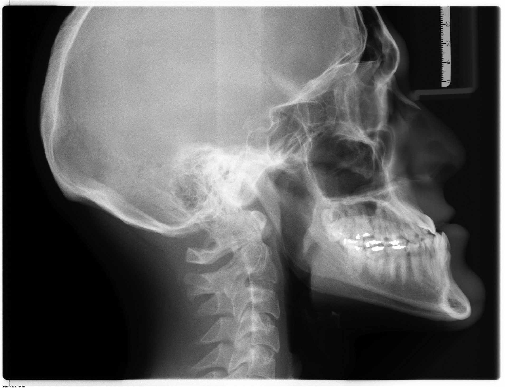
In the context of a dental X-ray, we are primarily concerned with stochastic effects rather than deterministic ones. Deterministic effects (like skin erythema or mucositis) require a high threshold dose that is never approached in diagnostic dentistry. Stochastic effects (such as carcinogenesis) do not have a threshold; theoretically, the probability of the effect increases with dose, but the severity does not. This is the biological basis for the ALARA principle (As Low As Reasonably Achievable). However, it is scientifically crucial to note that the effective dose of a modern digital intraoral radiograph is measured in microsieverts (µSv), often comparable to a few hours of natural background radiation from the sun and soil.
